
Page 77 - Understanding NCERT Science 09
P. 77
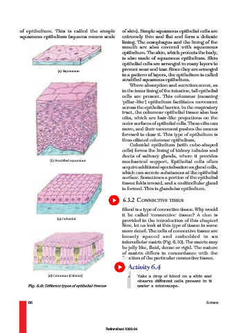
of epithelium. This is called the simple         of skin). Simple squamous epithelial cells are
              squamous epithelium (squama means scale          extremely thin and flat and form a delicate
                                                               lining. The oesophagus and the lining of the
                                                               mouth are also covered with squamous
                                                               epithelium. The skin, which protects the body,
                                                               is also made of squamous epithelium. Skin
                                                               epithelial cells are arranged in many layers to
                                                               prevent wear and tear. Since they are arranged
                                (a) Squamous
                                                               in a pattern of layers, the epithelium is called
                                                               stratified squamous epithelium.
                                                                  Where absorption and secretion occur, as
                                                               in the inner lining of the intestine, tall epithelial
                                                               cells are present. This columnar (meaning
                                                               ‘pillar-like’) epithelium facilitates movement
                                                               across the epithelial barrier. In the respiratory
                                                               tract, the columnar epithelial tissue also has
                                                               cilia, which are hair-like projections on the
                                                               outer surfaces of epithelial cells. These cilia can
                                                               move, and their movement pushes the mucus
                                                               forward to clear it. This type of epithelium is
                                                               thus ciliated columnar epithelium.
                                                                  Cuboidal epithelium (with cube-shaped
                                                               cells) forms the lining of kidney tubules and
                                                               ducts of salivary glands, where it provides
                            (b) Stratified squamous            mechanical support. Epithelial cells often
                                                               acquire additional specialisation as gland cells,
                                                               which can secrete substances at the epithelial
                                                               surface. Sometimes a portion of the epithelial
                                                               tissue folds inward, and a multicellular gland
                                                               is formed. This is glandular epithelium.


                                                               6.3.2 CONNECTIVE TISSUE
                                                               Blood is a type of connective tissue. Why would
                                                               it be called ‘connective’ tissue? A clue is
                                (c) Cuboidal
                                                               provided in the introduction of this chapter!
                                                               Now, let us look at this type of tissue in some
                                                               more detail. The cells of connective tissue are
                                                               loosely spaced and embedded in an
                                                               intercellular matrix (Fig. 6.10). The matrix may
                                                               be jelly like, fluid, dense or rigid. The nature
                                                               of matrix differs in concordance with the
                                                               function of the particular connective tissue.
                                                                 Activity 6.4

                           (d) Columnar (Ciliated)                    Take a drop of blood on a slide and
                                                                      observe different cells present in it
                  Fig. 6.9: Different types of epithelial tissues     under a microscope.


                  66                                                                                  SCIENCE


                                                       Rationalised 2023-24